Bones Basics
|
Introduction
Bones provide our bodies with the levers needed for movement, protection of the vital organs, important minerals for the blood, and
major cell types needed for defense against invaders (white blood cells) and oxygen for cellular respiration (red blood cells). Without bones, we will be crawling defenseless organisms. The various functions of bones, as
mentioned will mean that bones must be matched with complimentary features and structures to facilitate carrying out those functions, which in fact is the case, as we will see below.
Bones of the skeleton can be initially divided into 2 major classes,
those belonging to the axial skeleton and those belonging to the appendicular skeleton. The axial skeleton is comprised of bones that form
the long axis of the body. These include bones of the skull, vertebral column, and the thorax. The appendicular skeleton, in contrast, is comprised of
all the bones of the upper and lower limbs along with the shoulder and pectoral girdles, which attach the limbs to the axial skeleton. The structures of the bones that make
up both the axial and appendicular skeletons can be further divided into the following groups: 1. Long Bone: These bones are longer than they are wide. Some
examples include: the femur, phalanges, and humerous. 2. Irregular Bones: The name says it all. These bones are irregullarly shaped. Examples of this type
of bone include: the vertebrae and hip bones. 3. Flat Bones: These bones are flat and thin and often curved. Examples of these bones include: most skull bones, scapulae,
sternum, ribs. 4. Short Bones: These bones are roughly cubed shaped. Examples of short bones include: carpals and tarsals. Within the group short
bones falls the special bone type called Sesamoid Bones. These bones often form in a tendon. One example of this is the patella.
-O. James
© Orin James 2013
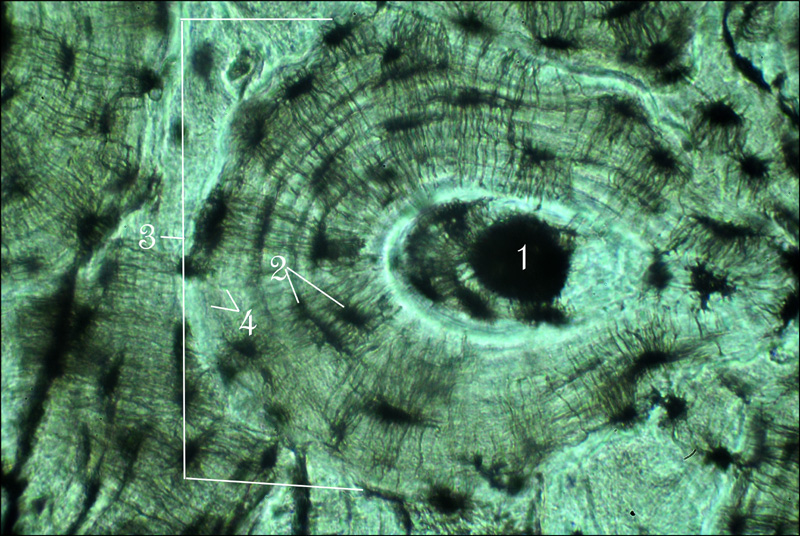
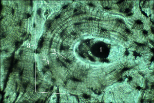 Microscopic Anatomy of Compact Bone Tissue
The image on the right is a photomicrograph of a cross section of bone tissue (400x).
(1) Central Canal (2) Lacunae containing osteocytes
(3) Osteon (4) Lamellae.
Click on image for larger representation. Photo by Orin James.
The appearance of compact bone tissue can be compared to a tree trunk that has been cut transversely, revealing the rings of age. If compared
to a tree the outermost ring will be the boundary of what is called the Osteon/Haversian System (3) of bone tissue. The osteon is the structural unit
of compact bone. Within it are hollow tubes of bone matrix, each being referred to as lamella (4). As we make our way towards the center of the osteon, one
notices that each matrix is riddled with Lacunae (2), which contains the osteocytes or bone cells. The osteocytes function to maintain the bone matrix
and act as stress sensors in cases of bone deformation or other stressful stimuli. At the center of the Osteon is the Central Canal (1), which serves
as the passageway for lymph vessels, blood vessels and nerves.
-O. James
© Orin James 2013
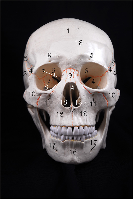
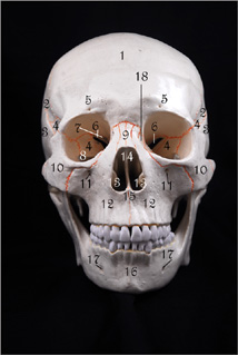 Bones of the Skull (Anterior View)
The image on the right is the anterior view of the skull. To view enlarged image in a new window, simply click on image. (image by Orin James).
The skull can be divided into two classes of bones, Cranial Bones and Facial Bones. The cranial bones can then be further divided into the Cranial Vault
and the Cranial Base. The cranial vault forms the superior, lateral and posterior aspects of the skull along with the forehead, while the cranial base
forms the skull's inferior aspect. The facial bones, a total of 14, comprise the bones of the face.
- Frontal Bone.
- Parietal Bones.
- Temporal Bones.
- Sphenoid Bone.
-
Supraorbital Foramen.
-
Superior Orbital Fissure.
-
Optic Canal.
- Inferior Orbital Fissure.
- Nasal Bone.
- Zygomatic Bone.
- Infraorbital Foramen.
- Maxilla.
- Inferior Nasal Conchae.
- Perpendicular Plate.
- Vomer.
- Mandible.
- Mental Foramen.
- Lacrimal Bone.
-O. James
© Orin James 2013
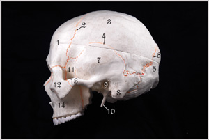 Bones of the Skull (Lateral View)
The image on the right is the lateral view of the skull, highlighting bones of the cranial vault. To view enlarged image in a new window, simply click on image. (image by Orin James).
Here are some of the bones of the cranium. A total of eight bones comprise the cranium, some of which are presented here. Please note that there are 2
parietal bones (one on each side), one frontal bone (forehead), one occipital bone (skull's posterior), two, temporal bones (one on each side, inferior to
parietal bones, a spenoid bone and an ethmoid bone, both which will be given special attention later in this page. the cranial bones join each
other at sutures.
- Frontal Bone (cranial vault).
- Coronal Suture - Runs across forehead.
- Parietal Bones - One on each side (cranial vault).
- Squamous Suture - One on each side.
- Occipital Bone (cranial vault).
- Lambdoid Suture.
- Temporal Bone - One on each side (cranial vault).
- Mastoid Process of Temporal Bone - One on each side.
- External Acoustic Meatus of Temporal Bone - One on each side.
- Styloid Process - One on each side.
- Sphenoid Bone.
- Zygomatic Bone - One on each side.
- Zygomatic Process - One on each side.
- Maxilla.
-O. James
© Orin James 2013
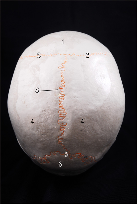
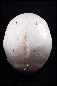 Bones of the Skull (Superior View)
The image on the right is the superior view of the skull, highlighting bones of the cranial vault. To view enlarged image in a new window,
simply click on image. (image by Orin James).
Here are some of the bones of the cranium. A total of eight bones comprise the cranium, some of which are presented here.
Please note that there are 2 parietal bones (one on each side), one frontal bone (forehead), one occipital bone (skull's posterior),
two, temporal bones (one on each side, inferior to parietal bones, a spenoid bone and an ethmoid bone, both which will be given
special attention later in this page. the cranial bones join each other at sutures.
- Frontal Bone (cranial vault).
- Coronal Suture.
- Sagittal Suture.
- Parietal Bones (cranial vault).
- Lambdoid Suture.
- Occipital Bone (cranial vault).
-O. James
© Orin James 2013
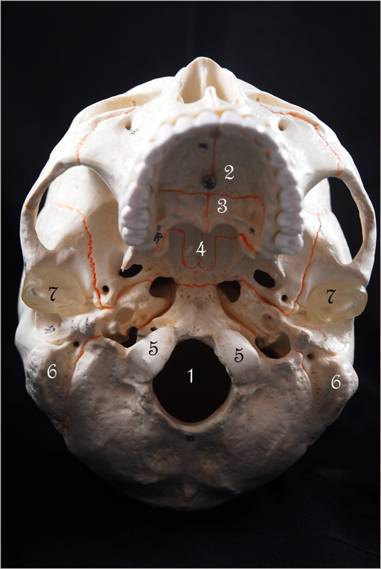
 Bones of the Skull (Inferior View)
The image on the right is the superior view of the skull, highlighting bones of the cranial vault. To view enlarged image in a new window,
simply click on image. (image by Orin James).
- Foramen Magnum.
- Palatine Process of Maxilla (roof of mouth).
- Palatine Bone.
- Vomer.
- Occipital Condyle.
- Mastoid Process.
- Mandibular Fossa.
-O. James
© Orin James 2013
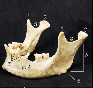 The Mandible
The image on the right is the mandible. (image by Orin James).
The Mandible is the largest facial bone and makes up the lower jaw and chin.
- Coronoid Process
- Mandibular Condyle (articulates with mandibular fossa).
- Mandibular Notch.
- Mental Foramen - One on each side.
- Ramus of Mandible.
- Mandibular Angle.
- Alveolar Margin (spaces between teeth).
-O. James
© Orin James 2013
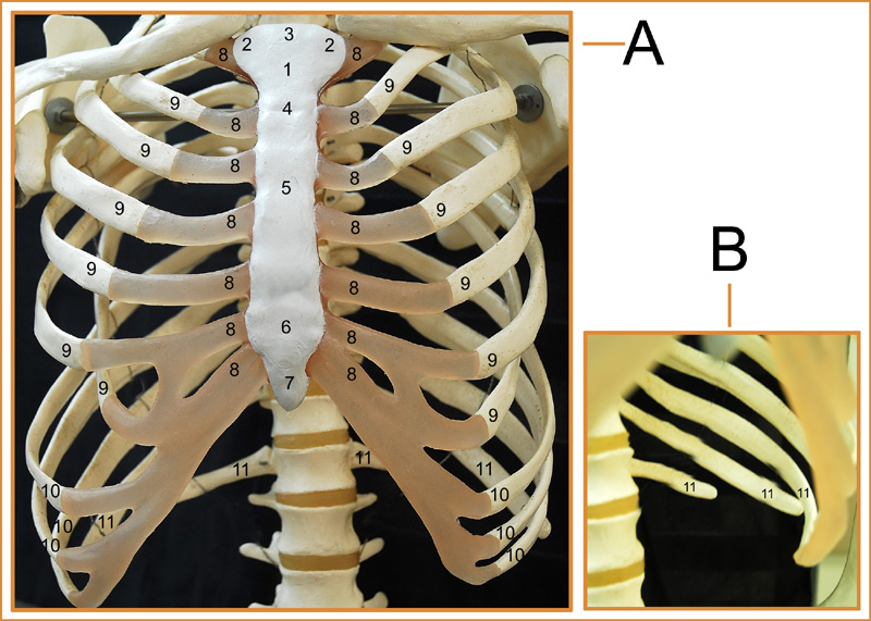
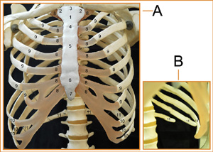 The Thoracic Cage (Anterior View)
The image on the right is the Thoracic Cage. (image by Orin James).
The thoracic cage provides the vital organs of the thoracic cavity (lungs, heart, blood vessels), with
protection.
Numbers 1-7 all comprise the sternum (breast bone).
- Manubrium of Sternal bone
- Clavicle Notch of Manubrium (Site where Clavicles articulate with Sternum).
- Jugular Notch of Manubrium.
- Sternal Angle (Here the Manubrium joins the Body of the Sternum).
- Body of Sternum.
- Xiphisternal Joint (site where body of Sternum joins the Xiphoid Process).
- Xiphoid Process.
- Costal Cartilage.
- True Ribs (There are 14 total, 7 on each side. These ribs connect directly to the Sternum)
- False Ribs (There are 10 total, 5 on each side. These ribes connect indirectly via the cartilages
of the superior true ribs. The lower 4 ribs, 2 on each side are called Floating Ribs, because
they do not connect at all to the thoracic cage.
-O. James
© Orin James 2013
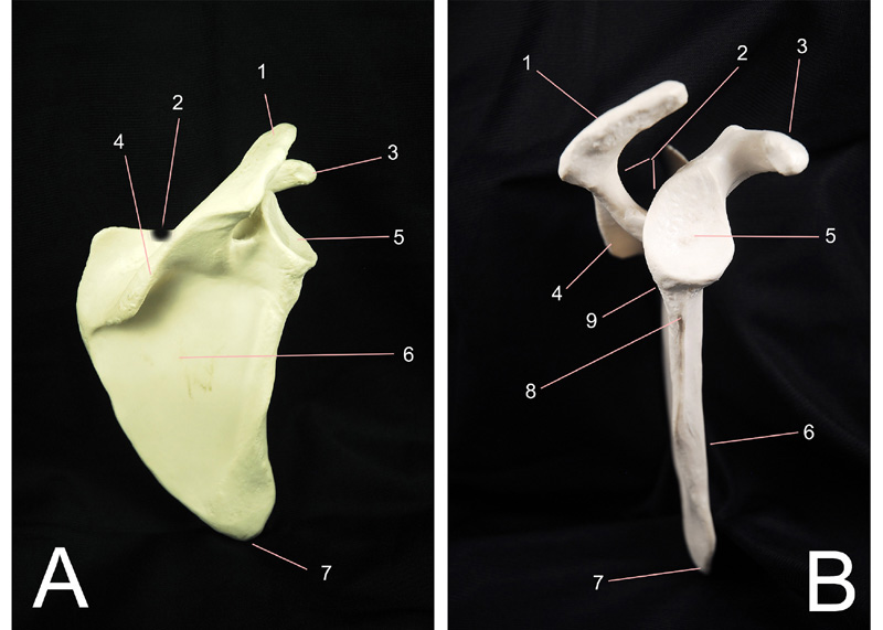
 The Scapula
The image on the right is that of the right scapula. Posterior aspect (A), lateral aspect (B). To see larger view, simply click on image. (images by Orin James).
The scapula, also known as the shoulder blade, is one of two bones that make up the pectoral girdles or shoulder girdles. The other bone is the clavicle. There is one pair of each bone,
one on the left and one on the right of the body. These girdles provide attachment of
the upper limbs to the axial skeleton. The pectoral girdles also provide sites of attachment for muscles responsible for moving the upper
limbs. These girdles are very light and allows great flexibility of the humerous. However, due to the light nature of this region, it is
highly susceptible to breaking, making shoulder dislocation very common.
- Acromion (articulates with the acromial end of clavicle)
- Suprascapular Notch (passage for nerves).
- Carocoid Process (helps anchor biceps to arm).
- Spine.
- Glenoid Cavity (loosly articulates with humerous bone).
- Infraspinous Fossa (A only, number 9 in B).
- Inferior Angle
- Infraglenoid Tubercle
- Infraspinous Fossa (B only, number 6 in A)
-O. James
© Orin James 2013
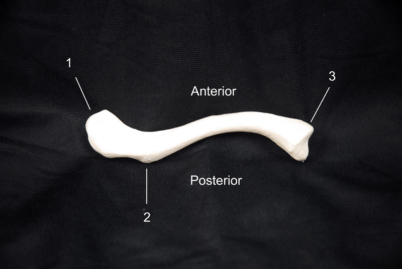
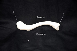 The Clavicle
The image on the right is the posterior view of the right clavicle. To see larger view, simply click on image. (images by Orin James).
The clavicle, also known as the collar bone, is one of two bones that make up the pectoral girdles or shoulder girdles. The other bone is the scapula. There is one pair of each bone,
one on the left and one on the right of the body. These girdles provide attachment of
the upper limbs to the axial skeleton. The pectoral girdles also provide sites of attachment for muscles responsible for moving the upper
limbs. These girdles are very light and allows great flexibility of the humerous. However, due to the light nature of this region, it is
highly susceptible to breaking, making shoulder dislocation very common. The clavicle is especially weak and often fractures when one attempts to
break a fall. Unfortuneatly, a posterior fracture of the clavicle may lead to damage of the subclavian artery.
- Acromial End (articulates with the scapula)
- Conoid Tubercle (site for ligament attachment that connects the clavicle to the scapula).
- Sternal End (articulates with sternal manubrium).
-O. James
© Orin James 2013
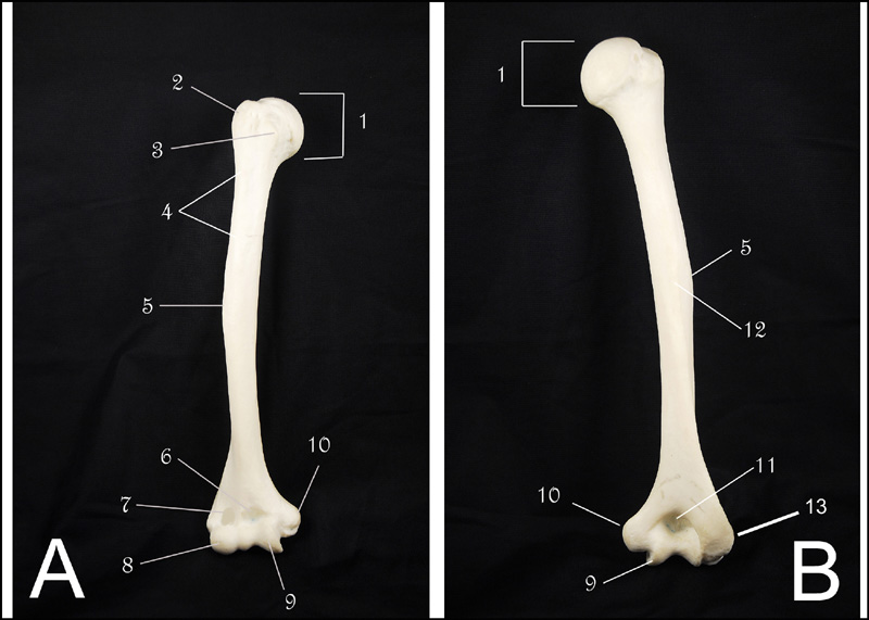
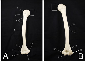 The Humerous
The image on the right shows the anterior view (A) and posterior view (B) of the humerous. To see larger view, simply click on image. (images by Orin James).
The humerous is the longest and largest bone of the upper limb. It articulates with the scapula (shoulder bone) and radius and ulna (forearm bones).
- Head of Humerous (articulates with the scapula)
- Greater Tubercle (site for muscle attachment).
- Lesser Tubercle (site for muscle attachment).
- Intertubercle Sulcus (guides a tendon of the biceps muscle of the arm to attachment point on glenoid cavity of scapula).
- Deltoid Tuberosity (site for deltoid muscle attachment).
- Coronoid Fossa (articulates with ulna for smooth elbow flexion).
- Radial Fossa (articulates with head of radius bone for smooth elbow flexion).
- Capitulum (articulates with radius).
- Trochlea (articulates with the trochlear notch of ulna).
- Medial Epicondyle (site for muscle attachment).
- Olecranon Fossa - POSTERIOR ONLY (articulates with olecranon process of ulna).
- Radial Groove - POSTERIOR ONLY (for course of radial nerve).
- Lateral Epicondyle.
-O. James
© Orin James 2013
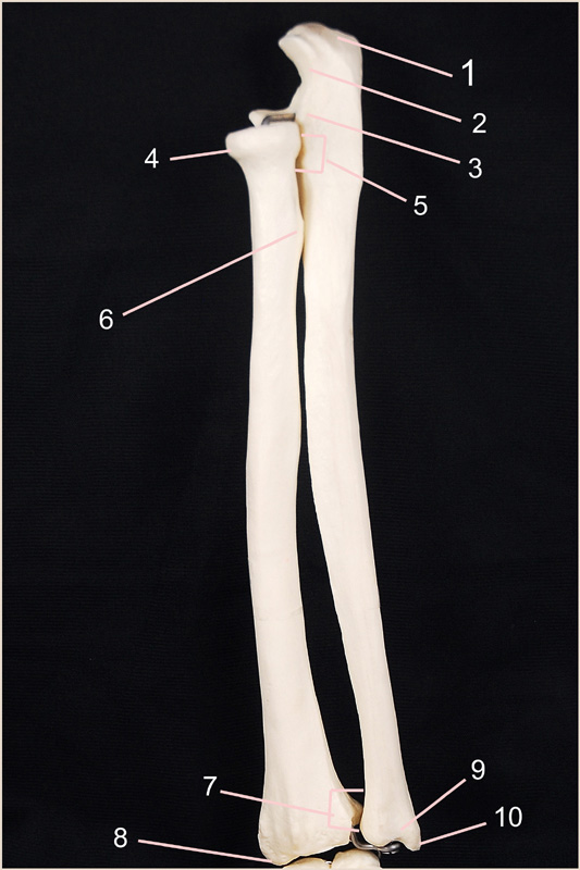
 The Radius and Ulna
The image on the right shows the anterior view of the radius (left) and ulna bones (right). To see larger view, simply click on image. (image by Orin James).
The radius and ulna make up the forearm or antebrachial region of upper limb. They are two parallel bones that articulate with the humerous and carpal bones.
- Olecranon Process (articulates olecranon fossa of humerous)
- Trochlear Notch (articulates/grips the trochlea of the humerous bone).
- Coronoid Process (along with trochlear notch, articulates/grips the trochlea of the humerous bone).
- Head of Radius (articulates with capitulum of humerous).
- Proximal Radioulnar Joint (site of proximal joint of ulna and radius).
- Radial Tuberosity (site of insertion for biceps muscle).
- Distal Radioulnar Joint (site of distal joint of ulna and radius).
- Radial Styloid Process (site of ligament attachments that run to the wrist).
- Head of Ulna.
- Ulnar Styloid Process (site of ligament attachments that run to the wrist).
-O. James
© Orin James 2013
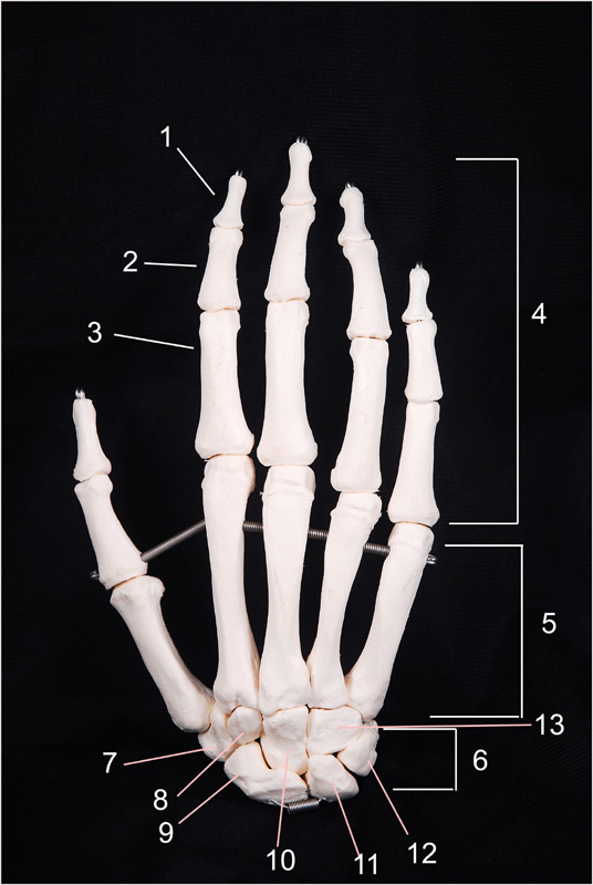
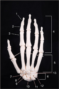 The Hand (Carpals and Phalanges)
The image on the right shows the posterior view of the right hand (palm facing down). To see larger view, simply click on image. (image by Orin James).
The hand is comprised of bones of: the carpus (wrist), metacarpus (palm) and phalanges (fingers). The carpals or wrist bones will
articulate with the radius and ulna bones.
- Distal Phalanx (plural = phalanges)
- Middle Phalanx (plural = phalanges)
- Proximal Phalanx (plural = phalanges).
- Phalanges (distal, middle, proximal) - THUMB DOES NOT HAVE A MIDDLE PHALANX.
- Metacarpals.
- carpals (wrist bones that articulate with distal radius and ulna bones).
- Trapezium.
- Trapezoid.
- Scaphoid.
- Capitate.
- Lunate.
- Triquetrum and Pisiform.
- Hamate.
-O. James
© Orin James 2013
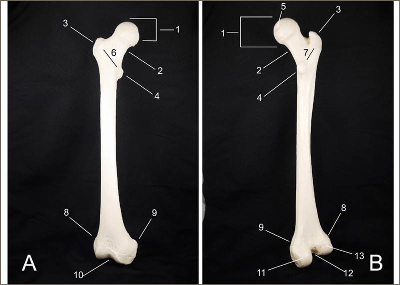
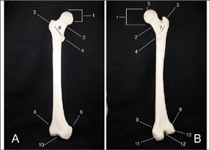 The Femur
The image on the right shows the anterior (A) and posterior (B) views of the right femur. To see larger view, simply click on image. (image by Orin James).
The femur is the strongest, largest, and longest bone in the body. It clearly bears most of the body's weight. Proximally, it articulates with the coxal bone
and distally with the tibia.
- Head of Femur (articulates with acetabulum of coxal bone).
- Neck of Femur.
- Greater Trochanter (site of muscle attachment for thigh and buttocks muscles).
- Lesser Trochanter (site of muscle attachment for thigh and buttocks muscles).
- Fovea Capitis (site of ligament attachment to acetabulum of coxal bone).
- Intertrochantric Line (A) - (connects the greater and lesser trochanters anteriorly).
- Intertrochantric Crest (B) - (connects the greater and lesser trochanters posteriorly).
- Lateral Epicondyle (site for muscle attachment).
- Medial Epicondyle (site for muscle attachment).
- Patellar Surface (A) - (site for articulation with patellar bone).
- Medial Condyle (B) - (site for muscle attachment).
- Intercondylar Fossa (B) - (articulates with intercondylar emminence and condyles of tibia).
- Lateral Condyle (B) - (site for muscle attachment).
-O. James
© Orin James 2013
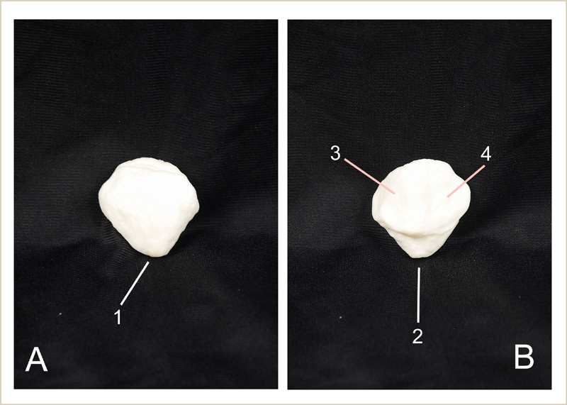
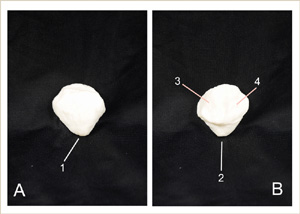 The Patella (Kneecap)
The image on the right shows the anterior (A) and posterior (B) views of the patella. To see larger view, simply click on image. (image by Orin James).
The patella functions to protect the knee joint anteriorly and enhance the leverage of the thigh
muscles across the knee. It is a sesamoind shaped bone that aids in securing the anterior thigh muscles
to the tibia bone.
- Apex of Patella.
- Surface for Patella Ligament.
- Facet for Medial Condyle of Femur.
- Facet for Lateral Condyle of Femur.
-O. James
© Orin James 2013
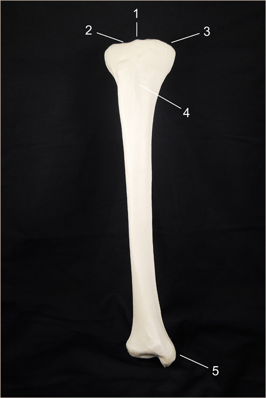
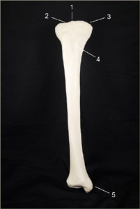 The Tibia
The image on the right shows the anterior view of the right tibia. To see larger view, simply click on image. (image by Orin James).
The tibia is the second strongest, largest, and longest bone in the body. It receives weight placed on the femur.
Proximally, it articulates with the femur bone and distally with the tarsals.
- Intercondylar Emminence (fits between intercondylar fossa of femur bone).
- Lateral Condyle (articulates with lateral condyle of femur bone).
- Medial Condyle (articulates with medial condyle of femur bone).
- Tibial Tuberosity (site of patellar ligament attachment).
- Medial Malleolus (forms the medial bulge of ankle).
-O. James
© Orin James 2013
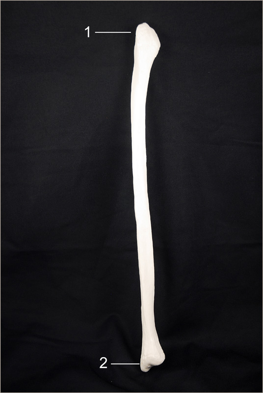
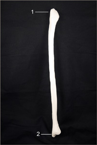 The Fibula
The image on the right depicts the anterior view of the right fibula. To see larger view, simply click on image. (image by Orin James).
The fibula articulates with the lateral aspects of the tibia. It does not bear weight, but muscles important for the leg
originate here.
- Head (articulates with lateral condyle of tibia).
- Lateral Malleolus (articulates with talus of foot and forms lateral bulge of ankle).
-O. James
© Orin James 2013
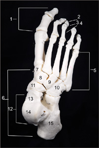
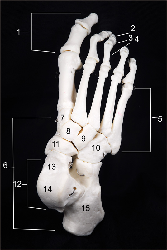
The image on the right shows the superior view of the right foot. To see larger view, simply click on image. (image by Orin James).
The foot is not only responsible for supporting our body weight, but also for propelling the entire body forward when walking or running.
- Phalanges.
- Distal Phalanx (plural = phalanges).
- Middle Phalanx (plural = phalanges).
- Proximal Phalanx (plural = phalanges).
- Metatarsals.
- Tarsals.
- Medial Cuneiform.
- Intermediate Cuneiform.
- Lateral Cuneiform.
- Cuboid.
- Navicular.
- Talus.
- Talus (articulates with tibia and fibula superiorly).
- Trochlea of Talus.
- Calcaneus (carries talus on superior surface and posteriorly attaches Achilles Tendon).
-O. James
© Orin James 2013
Physiological Mechanisms of Bone Formation/Modelling
|
Coming soon...
-O. James
© Orin James 2013