 Microscopic Anatomy of Skeletal Muscles
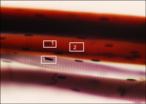
Image on right is a photomicrograph of a teased skeletal muscle fiber (150x, photo by Orin James).
Muscle tissue of humans exists in three forms, skeletal, smooth and cardiac.
Skeletal muscles are used to carry out voluntary actions and are controlled by the somatic division of
the nervous system. Because skeletal muscles are characterized by a striated appearance they are often
referred to as striped or striated muscles. Smooth muscles are used to carry out involuntary actions and
are controlled by the autonomic division of the nervous system. Unlike skeletal muscles,
smooth muscles do not appear striated. This is primarily due to the obvious lack of banding pattern found in
skeletal muscles.The last type of human muscle is the cardiac muscle. The cardiac muscle is used for the involuntary pumping
action of the heart and it too is controlled by the autonomic division of the nervous system.
Some of the characteristics of the cardiac muscle include the presence of intercalated discs and striations.
The remainder of this page will focus on skeletal muscles.
The image on your right is actually the second to smallest subunit (muscle fiber) of a skeletal muscle.
After one cuts through this subunit, one encounters the myofibril, which contains the contractile components.
However, before one reaches the muscle fiber, one is sure to see the epimysium, which wraps the belly of the
entire muscle and blends into deep fascia to help form tendons or sheet like aponeuroses that tether muscles to
bones. Beneath the epimysium are bundles called fascicles wrapped in perimysium.
Within each fascicle are the muscle fibers separated by endomysium and wrapped in sarcolemma.
Lastly, as indicated above, within each muscle fiber are myofibrils, which, again, contain the muscle’s
contractile units. The photomicrograph on the right shows a teased muscle fiber.
- Nuclei (1) - This dark oblong-shaped structure is the nucleus of the cell. As is apparent,
there are numerous nuclei on the muscle fiber.
- Bands (2) - The vertical light and dark banding pattern observed here is due to the perfect alignment of the
myofibrils. Along each myofibril are I bands (light) and A bands (dark). These bands produce a striated
appearance of the skeletal muscle, thereby giving it the epithet "striped muscle".
Within these bands are the contractile components of the muscle. More on contraction below.
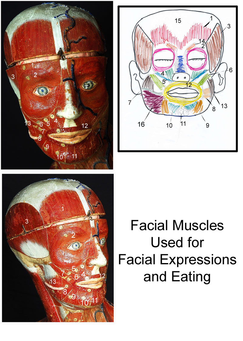
 Facial Muscles
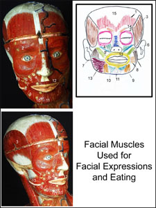
The images on the right represent the facial muscles of a human.
To view enlarged image in a new window, simply click on image (photos and drawing by Orin James).
As we begin the exploration of skeletal muscles, let's start with the superficial facial muscles.
These muscles are primarily used to communicate emotions and are involved in mastication (chewing). As you read the description of each
muscle, you will find that I use the terms: originate, and insert. The site of origin refers to the location where the muscle begins.
The site of insertion is usually on a different bone or muscle and is the location where that muscle terminates.
It is important to remember that as a muscle flexes/contracts, it will
move the bone or muscle from the site of insertion towards the site of origin.
- Epicranius Frontal Belly - This muscle of the forehead originates in the cranial aponeurosis (white portion posterior to muscle) and it inserts
in the the skin of the eyebrows and root of nose. It is responsible for raising the eyebrows. At the back of the cranium can be found the occipital
belly, which pulls the scalp posteriorly.
- Orbicularis Oculi - This muscle originates in the frontal and maxillary bones along with ligaments around the orbit. It inserts in the
tissue of the eyelid. Its function is to blink, squint and close the eye, along with pulling the eyebrows down inferiorly.
- Temporalis - This muscle originates in the temporal fossa and inserts in the coronoid process of the mandible. It is responsible for closing
the jaw.
- Levator Labii Superiosis - This muscle originates in the zygomatic and infraorbital margin of maxilla bones. It inserts in the skin and muscle of
the upper lip and the border of the nostril. It is responsible for opening the lip.
- Zygomaticus Minor - This muscle originates in the zygomatic bone and inserts in the skin at the corner of the mouth. It raises the lateral corners
of the mouth upward to smile :-)
- Zygomaticus Major - See zygomaticus minor.
- Risorius - This muscle originates in the fascia of the masseter muscle and inserts in the angle of the mouth. It is responsible for tensing
the lips and drawing the corner of the lips laterally.
- Buccinator - This muscle originates in the molar region of the maxilla and mandible bones. It inserts in the orbicularis oris. It is know as
the "trumpeteer" muscle, because it draws the corner of the the mouth laterally and compresses the cheeks as is observed when viewing a trumpet player
perform. It also holds food between the teeth during chewing.
- Depressor Anguli Oris - This muscle originates in the body of the mandible beneath the incisors teeth. It inserts in the skin and muscle
at an angle of the mouth below the insertion of the zygomaticus. It is responsible for drawing the mouth downward and laterally.
- Depressor Labii Inferiosis - This muscle originates in the body of the mandible slightly lateral to its midline. It inserts in the skin and
muscle of the lower lip. It is responsible for drawing the lower lip inferiorly.
- Mentalis - This muscle originates in the mandible below the incisors teeth. It inserts in the skin of the chin and is responsible for wrinkling
the chin.
- Orbicularis Oris - This circular muscle originates from the maxilla and mandible. Its fibers blend with fibers of other muscles associated
with the lips. It encircles the mouth and inserts into the muscles and skin at various angles fot the mouth.
- Masseter - This muscle originates in the zygomatic arch and maxilla. It inserts at the ramus and an angle of the mandible. It is responsible
for closing the jaw.
- Corrugator Supercilii - This muscle originates in the arch of the frontal bone above the nasal bone. It inserts in the skin of the eyebrow.
It is responsible for drawing the eyebrows medially and inferiorly.
- Galea Aponeurotica - This is not a muscle, but an aponeurosis used as the site of origin for the frontal and occipital bellies of the epicranius
muscle.
- Platysma - This muscle is certainly well known to individuals who shave from the mouth, down the neck. It originates in the fascia of the
chest and inserts in the lower end of the mandible, skin and the muscle at the corner of the mouth. It is responsible for tensing the skin of the
neck during shaving and giving that downward sag of the mouth appearance.
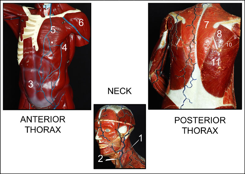
 Muscles of Thorax
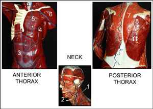
The collage on the right depicts a very small selection of anterior and posterior muscles of the thorax.
The center inset points to two muscles of the neck. To view enlarged image in a new window,
simply click on image (photos by Orin James).
- Sternocleidomastoid - This muscle of the neck originates in the manibrium of the sternum along with the medial
portion of the clavicle and it inserts in the the mastoid process of the temporal bone and the superior nuchal line
of the occipital bone. It is responsible for forward flexion of the neck and rotation of the head towards the shoulder
on opposite sides.
- Platysma - See facial muscles above (number 16).
- Abdominal Wall - Unfortunately, I do not have images of the other layers of the abdominal wall. Please note that
there are four muscular layers of the abdominal wall. These include the rectus abdominis (seen in image, this is the
six-pack layer, which many beach goers strive for), transversus abdominis (deepest muscle layer of the abdominal wall, fibers run horizontally),
external oblique (most superficial lateral muscle - this muscle is exercised when one does oblique curls.),
and the internal oblique (its fibers run perpendicular to the external oblique muscles).
- Rectus Abdominis - This layer originates in the pubic crest and symphysis and inserts in the xiphiod
process and the costal catilages of ribs number 5-7. It is responsible for rotating and flexing the vertebral column.
-
Transversus Abdominis - This layer originates in the inguinal ligament, iliac crest, the last five rib cartilages and lumber facia.
It inserts in the pubic crest and the linea alba. It is responsible for compressing abdominal contents.
-
External Oblique - This layer originates at the anterior surface of the last eight ribs. It inserts in the pubic
crest and tuberlces. It also inserts in the iliac crest and linea alba (white line).This layer is responsible
for rotating the trunk and flexing laterally.
-
Internal Oblique - This layer originates in the iliac crest and the inguinal ligament along with the lumbar fascia.
It inserts in the pubic crest and the cartilages of the last three ribs and the linea alba. This layer is responsible
for rotating the trunk and flexing laterally as the external oblique.
- Serratus Anterior - This muscle originates in the lateral aspect of the ribs numbers one through eight. It inserts
at the vertebral border of the anterior surface of the scapula. It is responsible for rotating the scapula and holding
the scapula against the chest wall.
- Pectoralis Major - This muscle originates in the clavicle, cartilage of ribs numbers one through six, the
sternum, and the aponeurosis fo the external oblique muscle. It inserts in the intertubercular sulcus of the
humerous in the form of a short tendon. It is responsible for rotating and flexing the arm along with adducting the arm medially.
- Deltoid - This muscle originates in the lateral one-third of the clavicle along with the spine of the clavicle
and acromion. It inserts in the deltoid tuberosity of the humerous. It is responsible for arm abduction.
- Trapezius - This muscle originates in the occipital bone, all the thoracic vertebrae, along with C7 and the
ligamentum nuchae. It inserts into the acromion and the spinous process of the scapula. It also inserts in the lateral
third of the clavicle. It is responsible for raising, rotating, retracting and stabilizing the scapula.
- Infraspinatus - This muscle originates in the infraspinous fossa of the scapula and inserts in the greater
tubercle of the humerous. It is responsible for lateral rotation of the humerous.
- Teres Minor - This muscle originates in the lateral margin of the scapula and inserts in the greater tubercle
of the humerous. Its function is the same as the infraspinatus.
- Teres Major - This muscle originates in the posterior surface at an inferior angle of the scapula.
It inserts in the intertubercular sulcus of the humerous. It is responsible for extending, medially rotating and adducting
the humerous.
- Latissimus Dorsi - This muscle originates indirectly as it attaches to the spinous processes of the lower
six thoracic vertebrae, lumbar vertebrae, iliac crest and last three to four ribs. It inserts at the floor of
the intertubercular sulcus of the humerous. It is responsible for extension, adduction and medial rotation of the
arm (this brings the arm down to provide a power stroke).
 Muscles of the Upper Arm
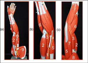
The collage on the right depicts a selection of arm muscles. (a) Medial view of left arm muscles. (b) closer view of
medial left arm muscles. (c) lateral view of left arm muscles. To view enlarged image in a new window,
simply click on image (photos by Orin James).
- Coracobrachialis - This muscle originates in the coracoid process of the scapula and inserts in the medial surface
of the humerous shaft. It is responsible for flexing and adducting the humerous.
- Biceps Brachii - The name biceps refer to the number of heads the muscle has. In this case
there are two heads, a short one and a long one. The short head originates at the coracoid process. The tendon
of the long head runs in the intertubercular sulcus of the humerous and within the capsule of the shoulder joint.
It inserts into the radial tuberosity of the radius bone. It is responsible for flexion of the elbow and supination
of the forearm. This is also know as the muscle that "turns the corkscrew and pulls the cork".
- Brachialis - This muscle is deep to the biceps brachii and originates in the distal portion of the anterior region of the humerous. It inserts
in the coronoid process of the ulna bone. It is responsible for flexing the forearm.
- Medial Head of Triceps Brachii - (a) & (b) only. Again, triceps refer to the number of heads the muscle has.
In this case, there are three heads for this muscle. Number four depicts the medial head of the triceps brachii.
The medial head originates in the distal radial groove on the posterior region of the humerous and inserts in the
olecranon process of the ulna bone. This muscle as a whole is a relatively powerful forearm extensor. It is also
an antagonist of the biceps brachii and brachialis muscles (see 5).
- Long Head of Triceps Brachii - Again, triceps refer to the number of heads the muscle has.
In this case, there are three heads for this muscle. Number five depicts the long head of the triceps brachii.
The long head originates in the posterior region of the humerous and inserts in the
olecranon process of the ulna bone. This muscle as a whole is a relatively powerful forearm extensor. It is also
an antagonist of the biceps brachii and brachialis muscles (see 7).
- Brachioradialis - This muscle originates in the lateral ridge at the distal end of the humerous and inserts
at the base of the styloid process ot the radius bone. It functions as a synergistic muscle for flexing the forearm.
- Lateral Head of Triceps Brachii - Again, triceps refer to the number of heads the muscle has.
In this case, there are three heads for this muscle. Number seven depicts the lateral head of the triceps brachii.
The lateral head originates inferior margin of the glenoid cavity of the scapula and inserts in the
olecranon process of the ulna bone. This muscle as a whole is a relatively powerful forearm extensor. It is also
an antagonist of the biceps brachii and brachialis muscles (see 4 & 5)
- Deltoid - See muscles of thorax above.
- Infraspinatus & Supraspinatus - (Infraspinatus) This muscle originates in the infraspinous fossa of the scapula and inserts in the greater
tubercle of the humerous. It is responsible for lateral rotation of the humerous. (Supraspinatus) This muscle originates
in the supraspinous fossa of the scapula and inserts in the greater tubercle of the humerous. It is responsible for
initiating the abduction of the humerous and stabilizing the shoulder joint.
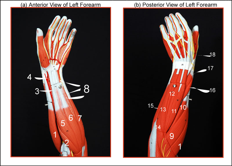
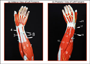 Muscles of the Forearm
The collage on the right depicts a selection of arm muscles. (a) Anterior view of left forearm muscles. (b) Posterior
view of left forearm muscles. To view enlarged image in a new window,
simply click on image (photos by Orin James).
- Brachioradialis - See number six of "muscles of arm" above.
- Pronator Teres - This muscle originates in the medial epicondyle of the humerous and coronoid process of the
ulna. It inserts in the midshaft of the radius. It is responsible for pronation of the forearm.
- Flexor Pollicis Longus - It is very difficult to clearly demarcate in this image, but know that it is a deep
anterior forearm muscle. It originates in the anterior surface of the radius and its interosseous membrane. It inserts
in the distal phalanx of the thumb. It is responsible for flexing the thumb.
- Pronator Quadratus - Another deep muscle. It is the deepest muscle of the forearm. it originates at the distal
end of the the anterior region of the ulna bone. It inserts at the distal anterior region of the radius. It is responsible
for pronating the arm.
- Flexor Carpi Radialis - This muscle originates on the medial epicondyle of the humerous and inserts in the base of
metacarpals 2 and 3. It is responsible for flexing the wrist and abducting the hand.
- Palmaris Longus - This muscle originates in the medial epicondyle of the humerous and inserts in the palmar
aponeurosis, along with the skin and fascia of the palm. it is responsible for tensing the skin and the fascia of
of the arm.
- Flexor Carpi Ulnaris - This muscle originates in the medial epicondyle of the humerous and the olecranon
process along with the posterior region of the ulna. It inserts in the base of metacarpal 5 and the pisiform and
hamate carpal bones. It is responsible for flexing the wrist and adducting the hand.
- Flexor Digitorum Superficialis- The image shows three lines pointing to number eight. However, it is one muscle
,which is deep to the palmaris longus, flexor carpi radialis, and the flexor carpi ulnaris. It originates in the medial epicondyle of the humerous,
the coronoid process of the ulna and the shaft of the radius. They insert into the middle phalanges of the fingers 2-5. It is responsible for flexing the wrist, and the middle
phalanges of the fingers 2-5.
- Extensor Carpi Radialis Longus - This muscle originates in the lateral supracondylar ridge of the humerous and
inserts at the base of metacarpal 2. It is responsible for abducting the wrist.
- Extensor Carpi Radialis Brevis - This muscle originates in the lateral epicondyle of the humerous and inserts at the base of metacarpal 3.
It is responsible for abducting and extending the wrist.
- Extensor Digitorum - This muscle originates in the lateral epicondyle of the humerous and inserts via
four tendons into the distal phalanges of fingers 2-5. It is responsible for extending the fingers.
- Extensor Digit Minimi -
- Extensor Carpi Ulnaris - This muscle originates in the lateral epicondyle of the humerous and posterior border
of the ulna. It inserts in the base of metacarpal 5. It is responsible for extending and adducting the wrist.
- Anconeus - This muscle originates in the lateral epicondyle of the humerous and inserts into the lateral aspect
of the olecranon process of the ulna. It is responsible for abducting the ulna while the forearm pronates.
- Flexor Carpi Ulnaris - See number 7.
- Abductor Pollicis Longus - This is a deep muscle and runs parallel to the extensor pollicis longus. It originates
at the posterior region of both the radius and ulna. It inserts at metacarpal 1 and the trapezium wrist carpal.
- Extensor Pollicis Brevis - This muscle is deep to the extensor carpi ulnaris. It originates in the dorsal shaft
of both the ulna and radius. It inserts at the base of the proximal phalanx of the thumb (brevis).
It is responsible for extending the thumb.
- Extensor Pollicis Longus - This muscle is deep to the extensor carpi ulnaris. It originates in the dorsal shaft
of both the ulna and radius. It inserts at the base of the distal phalanx of the thumb (longus).
It is responsible for extending the thumb.
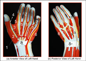 The Hand
The collage on the right depicts a selection of the left hand muscles and tendons.
(a) Anterior view of left hand muscles and tendons. (b) Posterior
view of left hand muscles and tendons. To view enlarged image in a new window,
simply click on image (photos by Orin James).
- Thenar Muscles of the Thumb (1-3, see special note) - These muscles represent the ball of the thumb and they are divided into 4 muscles.
SPECIAL NOTE: The muscle Opponens Pollicis, which is also a member of this group is not shown.
The opponens pollicis originates the flexor retinaculum and trapezium carpal. It inserts on the whole anterior
side of metacarpal I. Number 1: represents the Abductor Pollicis Brevis, which originates in the flexor retinaculum and surrounding carpals.
It inserts in lateral base of the thumbs proximal phalanx. It is responsible for abducting the thumb.
- Flexor pollicis Brevis _ This muscle is also part of the thenar muscles that make up the ball of the thumb.
It originates in the flexor retinaculum and surrounding carpals. It inserts in the lateral side of the base
of the proximal phalanx of the thumb. It is responsible for flexing the thumb.
- Adductor Pollicis - This muscle is also part of the thenar muscles that make up the ball of the thumb.
It originates in the capitate bone and at the bases of metacarpals II through IV and the front of metacarpals III.
It inserts into the medial side of the base of the proximal phalanx of the thumb. It is responsible for adducting
the thumb and helping to oppose the thumb.
- Tendon of Flexor Pollicis Longus.
- Lumbricals - There are four of these muscles, one for each finger, except the thumb. They originate on the
lateral side of each tendon of the flexor digitorum profundus (no image available yet) in the palm. They insert on the lateral edge of
the extensor expansion on the proximal phalanx of fingers 2-5. Their origins (from the tendons of another muscle)
make them unusual muscles.
- Hypothenar Muscles in the Ball of Little Finger - Like the thenar muscles of the thumb, here we have three
separate muscles (see special note). This image only shows you the Abductor Digiti Minimi Number 6, which originates in the pisiform carpal
and inserts on the medial side of the proximal phalanx of the little finger. It is responsible for abducting the
little finger at the metacarpophalangeal joint. SPECIAL NOTE: Deep to the abductor digiti minimi is the opponens
digiti minimi (not shown), which originates at the hamate carpal and flexor retinaculum. It inserts along the medial
side of metacarpal V. The last and of the hypothenar muscles is the flexor digiti minimi brevis. It originates in the
hamate carpal and flexor retinaculum. It is characterized as the lateral deep muscle of the hypothenar group. It
inserts in the medial side of the the proximal phalanx of the little finger. It is responsible for flexing the middle
finger at the metacapophalangeal joint.
- Tendons of Flexor Digitorum Superficialis - See number 8 of forearm muscles above.
- Flexor Retinaculum - This wristband-like thick fascia is responsible for securing all the tendons at the wrist.
- Tendons of Extensor Digitorum - See number 11 of forearm muscles above.
- Extensor Expansion -
- Interossei - There are both dorsal and palmar interossei. These muscles fill the spaces between the
metacarpals. They originate at the sides of the metacarpals and insert at the extensor expansion. The palmar
inerossei will adduct the fingers, while the dorsal interossei abduct the fingers.
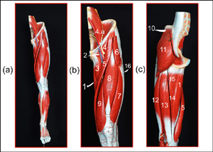 Muscles of the Thigh
The collage on the right depicts a selection of muscles of the left leg.
(a) Anterior view of muscles of left leg. (b) Anterior view of left thigh. (c) Posterior view of left thigh (at
a slight angle showing slight medial view).
To view enlarged image in a new window,
simply click on image (photos by Orin James).
- Gracilis - This muscle originates in the inferior ramus and body of the pubis. It inserts at the medial region
of the tibia, slightly inferior to the medial condyle. It is responsible for adducting the thigh, flexing and
rotating the thigh medially.
- Pectineus - This muscle originates at the pectineal line of the pubis and inserts inferiorly from the lesser
trochanter to the linea aspera of the femur. It is responsible for adducting, flexing and rotating the thigh medially.
- Adductor longus - This muscle originates at the pubis, near the the pubic symphysis. It inserts at the linea apsera
of the femur.
- Iliopsoas - This muscle is further divided into the iliacus and psoas major muscles. The iliacus originates
in the iliac fossa and crest, along with the lateral sacrum. It inserts on and slightly below the lesser trochanter
of the femur. The psoas major originates on the transverse processes, bodies and discs of the T12 vertebra, and the
remaining lumbar vertebrae (4.a includes parts of psoas major, minor and the Quadratus lumborum). Both these muscles are responsible for flexing the trunk on the thigh, flexing the thigh
and lateral flexion of the vertebral column.
- Sartorius - Named after a famous Roman tailor, this muscle is also known as the "tailor's muscle". It is
responsible for placing the legs in a cross-legged position. It is also the longest muscle of the body.
It originates at the anterior superior region of the
iliac spine and inserts via an aponeurosis into the medial surface of the proximal tibia.
- Tensor Fasciae Latae - This muscle originates at the anterior aspect of the iliac crest and the anterior
superior iliac spine. It inserts in the iliotibial tract (number 16). It is responsible for abduction and medial rotation
of the thigh.
- Vastus Lateralis - This is one of the four muscles that make up the Quadriceps Femoris, along
with the femoris rectus, vastus medialis (8 & 9) and vastus intermedius (not shown). It originates
at the greater trochanter, intertrochanteric line and linea aspera of the femur. It inserts at the tibial tuberosity
and patella. It is responsible for extending and stabilizing the knee.SPECIAL NOTE:
The vastus intermedius also make up the quadriceps femoris. The vastus intermedius
is not shown as it is obsured by the rectus femoris. However, it originates in the anterior and lateral surface
of the femur and inserts into the tibial tuberosity and patella. It is responsible for extending the knee.
- Rectus Femoris - This is one of the four muscles that make up the Quadriceps Femoris, along with
the vastus lateralis, vastus medialis (7 & 9) and vastus intermedius (not shown). It originates at
the anterior inferior iliac spine and the superior margin of the acetebelum. It inserts into the tibial tuberosity
and patella. It is responsible for extending the knee and flexing the thigh at the hip.SPECIAL NOTE:
The vastus intermedius also make up the quadriceps femoris. The vastus intermedius
is not shown as it is obsured by the rectus femoris. However, it originates in the anterior and lateral surface
of the femur and inserts into the tibial tuberosity and patella. It is responsible for extending the knee.
- Vastus Medialis - This is one of the four muscles that make up the Quadriceps Femoris, along with
the vastus lateralis, femoris rectus (7 & 8) and vastus intermedius (not shown). It originates in the linea aspera and intertrochanteric line of the
femur. It inserts at the tibial tuberosity and patella. It is responsible for extending the knee and stabilizing
the patella.SPECIAL NOTE:
The vastus intermedius also make up the quadriceps femoris. The vastus intermedius
is not shown as it is obsured by the rectus femoris. However, it originates in the anterior and lateral surface
of the femur and inserts into the tibial tuberosity and patella. It is responsible for extending the knee.
- Gluteus Medius - This muscle originates at the upper lateral surface of the ilium. It inserts at the greater
trochanter of the femur. It is responsible for abducting and rotating the thigh medially.
- Gluteus Maximus - This muscle originates in the dorsal region of the ilium, sacrum and coccyx. It inserts into
the gluteal tuberosity of the femur and the iliotibial tract (number 16).
- Biceps Femoris - This muscle resides in the posterior compartment of the thigh and is one of three that make up
the Hamstrings, along with the semitendinosus and the semimembranosus (13 & 15). As the name implies,
it has two heads, a long and a short head. The long head originates in the ischial tuberosity and inserts into the head
of the fibula and lateral condyle of the tibia. The short head originates in the linea aspera and distal femur and
inserts into the head of the fibula and the lateral condyle of the tibia. This muscle is responsible for extending
the thigh and flexing the knee.
- Semitendinosus - This muscle resides in the posterior compartment of the thigh and is one of three that make up
the Hamstrings, along with the biceps femoris and the semimembranosus (12 & 15). This muscle originates in
the ischial tuberosity and inserts into the medial aspect of the upper tibial shaft. It is responsible for extending
the thigh and flexing the knee.
- semimembranosus - This muscle resides in the posterior compartment of the thigh and is one of three that make up
the Hamstrings, along with the biceps femoris and the semitendinosus (12 & 13). This muscle originates in
the ischial tuberosity and inserts at the medial condyle of the tibia and the lateral condyle of the femur. It is
responsible for extending the thigh and flexing the knee.
- Adductor Magnus - This muscle originates in the ischial and pubic rami and the ischial tuberosity. It inserts
into the linea aspera and adductor tubercle of the femur. It is responsible for adducting, flexing and medially
rotating the thigh.
- Iliotibial Tract - This thick fascia ensheaths all the muscles of the thigh.
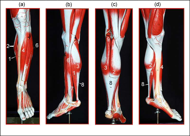
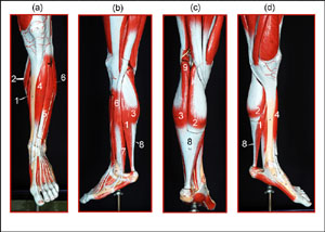 Muscles of the Lower Leg
The collage on the right depicts a selection of muscles of the left leg.
(a) Anterior view of muscles of left leg. (b) lateral view of left leg. (c) Posterior view of left leg. (d)
Medial view of left leg. To view enlarged image in a new window,
simply click on image (photos by Orin James).
- Soleus - This muscle originates in the proximal region of the tibia and fibula bones. It inserts into the calconeus
by way of the calconeal tendon (number 8). It is responsible for flexing the plantar.
- Gastrocnemius (medial head) - This muscle has two heads that originate from the medial and lateral condyles
of the femur bone. Both insert into the calcaneus by way of the calcaneal tendon (number 8). This muscle is responsible
for plantar flexion at the ankle.
- Gastrocnemius (lateral head) - This muscle has two heads that originate from the medial and lateral condyles
of the femur bone. Both insert into the calcaneus by way of the calcaneal tendon (number 8). This muscle is responsible
for plantar flexion at the ankle.
- Tibialis Anterior - This muscle originates in the lateral condyle and upper two-thirds of the tibia and
interosseous membrane. It inserts into the inferior surface of the first cuneiform and metatarsal 1 of the foot.
It is responsible for dorsiflexion.
- Extensor Digitorum Longus (?) - This muscle originates in the lateral condyle of the tibia and the proximal
three-fourths of the fibula and interosseous membrane. It inserts into the middle and distal phalanges of toes
2-5 via a tendon, which divides into four parts. It is responsible for flexing the toes.
- Fibularis Longus - This muscles originates at the head and upper portion of the fibula. It inserts by a long
tendon under the foot to metatarsal 1 and the medial cuneiform. It is responsible for everting the foot and flexing
the plantar.
- Fibularis Brevis - This muscle originates in the distal region of the the fibula's shaft. It inserts by a
tendon running behind the lateral malleolus to insert on the proximal end of metatarsal 5. It is responsible for
everting the foot and flexing the plantar.
- Calcaneal Tendon-
- Popliteus - This muscle originates in the lateral condyle of the femur and lateral meniscus. It inserts into
the proximal tibia. It is responsible for unlocking the knee when flexing the knee occurs, by rotating and flexing
the leg medially.
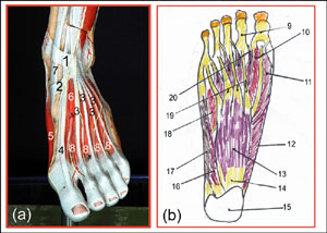
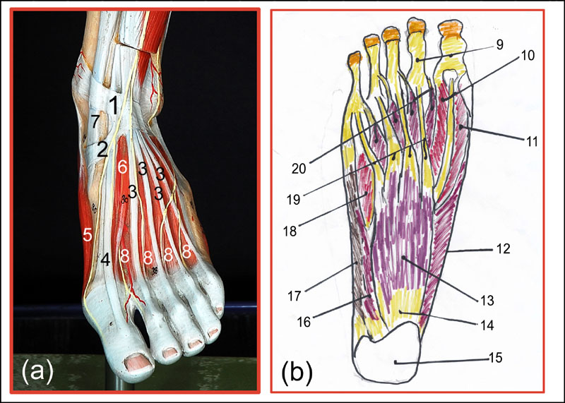
The collage on the right depicts a selection of muscles of the left foot and right foot (plantar view).
(a) Anterior view of tendons and muscles of left foot. (b) Drawing of the plantar view of the right foot.
To view enlarged image in a new window,
simply click on image (photo and drawing by Orin James).
- Superior Extensor Retinaculum - This leg brace-like tissue stabilizes the tendons for the extensor digitorum
longus (see number 5 of lower leg muscles).
- Inferior Extensor Retinaculum - See Superior Extensor Retinaculum.
- Tendons of Extensor Digitorum Longus - See number 5 of lower leg muscles above.
- Tendon of Extensor Hallucis Longus - Image of muscle not shown.
- Abductor Hallucis - This muscle originates on the tuberosity on the inferior surface of the calcaneus and
inserts on the medial side of the proximal phalanx of the big toe. It is responsible for abduction at the
metatarsophalangeal joint of the big toe.
- Extensor Hallucis Brevis - This muscle originates at the superior surface of the anterior calcaneus.
It inserts on the dorsal surface of the base of the proximal phalanx of the big toe.
- Tendon of Tibailis Anterior - See number 4 of lower leg muscles above.
- Dorsal Interossei - These muscles (4) originate on the sides of the metatarsal bones and insert on the
medial and lateral sides of the second toe and on the lateral sides of the third and fourth toes.
SPECIAL NOTE: Numbers 9-20 represent the tendons and muscles of the plantar view (from the bottom)
of the RIGHT foot!
- Fibrous Tendon Sheaths -
- Flexor Hallucis Brevis - This muscle originates in the cuboid and lateral cuneiform bones. It inserts into the
proximal phalanx of the big toe. It is responsible for flexion at the metatarsophalangeal joint of the big toe.
- Flexor Hallucis Brevis - See number 10.
- Abductor Hallucis - See number 5.
- Flexor Digitorum Brevis - This muscle originates at the inferior region the tuberosity of the calcaneus.
It inserts at the sides of the middle phalanges of toes 2-5. It is responsible for flexion at the the proximal
interphalangeal joints of toes 2-5.
- Plantar Aponeurosis -
- Calcaneus -
- Abductor Digiti Minimi - This muscle originates in the calcaneal tuberosity and inserts on the lateral side
of the base of the little toe's proximal phalanx. It is responsible for abducting and flexing the little toe.
- Abductor Digiti Minimi - See number 16.
- Flexor Digiti Minimi Brevis - This muscle originates at the base of the metatarsal bone IV. It inserts on the
lateral side of the proximal phalanx of toe 5. It is responsible for flexing the little toe at the metatarsophalangeal
joint.
- Tendons of Flexor Digitorum Brevis -
- Lumbricals- These muscles (4) originate at the tendons of the flexor digitorum longus (not shown) and insert
at the insertions of the extensor digitorum longus. They are responsible for flexing the toes at the metatarophalangeal
joints and extending the toes at the interphalangeal joints.
Skeletal Muscle Contraction
|
Coming soon...
-O. James
© Orin James 2012
 Muscles of Thorax
 Muscles of the Upper Arm
 Muscles of the Forearm
 The Hand
 Muscles of the Thigh
 Muscles of the Lower Leg
 Tendons and Muscles of the Foot